
“Glasses-free 3D allows us to directly visualize tumor and organ relationships, making treatment planning more efficient and consistent across the team.”
— Radiation Oncology Team, Hualien Tzu Chi Hospital
The Spatial Interpretation Challenge in Radiation Therapy
In radiationtherapy, precision is critical. Clinicians must deliver an effective dose to the tumor while protecting surrounding healthy tissue. That requires aclear, three-dimensional picture of the patient's anatomy.
However, current treatment planning still relies heavily on 2D cross-sectional images. Clinicians must mentally reconstruct complex 3D spatial relationships from numerous CT and MRI slices. Physicians trace tumor boundaries slice by slice, while physicists cross-check dozens of images to ensure full target coverage without compromising critical organs. For complex cases, particularly those nearcritical structures, this "inferring 3D from 2D" process imposes a significant cognitive burden, making interpretation more difficult and often prolonging interdisciplinary discussions.
To address this limitation, we collaborated with Hualien Tzu Chi Hospital to build the world’sfirst glasses-free 3D Smart Educational Training and Decision Support Platform for Radiation Oncology Treatment, enabling clinicians to see what they previously had to imagine.
The Real-World Impact on Clinical Workflow
Before adopting our technology, theradiotherapy team faced the following bottlenecks:
Overall, the spatial understanding gapinherent in 2D imaging not only increases clinical workload but may also affectthe efficiency of treatment plan review and the quality of cross-teamcommunication.
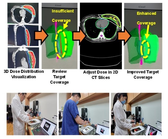
Solution: Lixel SpatiumNEX RTSS Glasses-Free 3D SmartEducational Training and Decision Support Platform
Hualien Tzu ChiHospital adopted the Lixel SpatiumNEX RTSS Platform, which convertsradiation therapy plans into intuitive 3D visualizations. This enablesclinical teams to quickly understand the spatial relationships amongtumors, organs at risk, and dose distributions.
Core features of the platform include:
Results: Measurable Gains in Speed, Consistency, and Safety
After implementing the glasses-free 3Dplatform, clinical data showed significant efficiency and safety improvements:
✅ Faster planning. Lessexperience dependency.
● 50% reduction in planningtime: Average review time shortened by half,greatly accelerating clinical workflows.
● 76% improvement inconsistency: The time gap between senior and juniorphysicists dropped from 67 minutes to just 16 minutes, significantly improvingplanning consistency.
✅ Fewer blind spots. Greaterpatient safety.
● Precise tumor coverage: Ensures full dose coverage of tumor margins, reducing recurrencerisk.
● Critical tissue protectionverified: Clearly confirms that adjacent tissuesare within safe limits, effectively preventing potential treatment blind spotsand enhancing overall safety.

Conclusion: Innovative Technology Achieves NationalRecognition
Withglasses-free 3D visualization, the Radiation Oncology team at Hualien Tzu ChiHospital has moved beyond the limitations of traditional 2D imaging. Clinicaldecision-making is no longer dependent on mental reconstruction, but groundedin direct spatial perception of tumors, organs at risk, and dose distributions.
This shiftnot only improves efficiency and consistency, but fundamentally changes howclinical teams interpret and communicate spatial information—reducing cognitive burden and enabling more aligned, confidentdecision-making across disciplines.
This technologyhas been awarded the National Innovation Award – Clinical InnovationCategory, and was selected for presentation at NVIDIA GTC 2026 Poster,showcasing Taiwan’s innovative integration of spatial computing into precisionmedicine on the international stage.
